Our patients rate us 4.9 out of 5 — 118 Google reviews

Around 25’000 refractive procedures2’500–3’000 laser procedures a year
Since 2006 in refractive surgeryevery laser procedure performed personally by Dr. Derhartunian

Read all reviews →

In brief

What is keratoconus?

Keratoconus is a disease of the cornea: it becomes thinner and bulges forward in a cone shape, and vision becomes distorted. It usually begins in adolescence and affects about 1 in 375 people. At EyeLaser Zürich, corneal crosslinking (CXL) stops the progression; for progressive keratoconus, basic health insurance pays, and as a self-payer it costs CHF 2’300 per eye.

Keratoconus at a glance — EyeLaser Zürich
Medical term Keratoconus (conical cornea); ICD-10 H18.6
Frequency about 1 in 375 people; diagnosed at an average age of 28, more common in men (Godefrooij et al. 2017)
Typical signs distorted, blurred vision, rapidly changing glasses prescriptions, double images in one eye, glare
Assessment Corneal tomography with the MS-39 AL, repeated after a few months to measure progression
Treatments Crosslinking (stops progression) · CAIRS, topography-guided laser, ICL (improve vision) · rigid contact lenses · corneal transplant as a last resort
Health insurance Crosslinking for progressive keratoconus: mandatory benefit of basic health insurance (KLV Annex 1). We nevertheless recommend obtaining a cost approval before the procedure and will support you with this.

How can you recognise keratoconus, and how does it develop?

Trifokallinsen

In keratoconus, the normally round cornea becomes thinner and bulges forward in a cone shape. The irregular shape distorts the image. The condition usually begins in puberty and often progresses until the age of 30 to 40; as a rule, both eyes are affected, often to different degrees.

Signs

  • Blurred or distorted vision that can no longer be fully corrected with glasses
  • Glasses prescriptions, especially the astigmatism, change within a short time
  • Double or multiple images in one eye, glare and halos at night

Who is at risk?

In an analysis of 29 studies, the risk was around six times higher with keratoconus in the family and around three times higher with frequent eye rubbing; allergies, asthma and eczema were also more common (Hashemi et al. 2020). You should therefore avoid rubbing your eyes.

When to see an ophthalmologist immediately? If an eye with keratoconus suddenly becomes cloudy and painful, fluid may have been forced into the cornea (acute hydrops). Have this checked by an ophthalmologist on the same day; at night and at weekends via the emergency department of the hospital on duty.
How is keratoconus diagnosed, and when is it progressing?

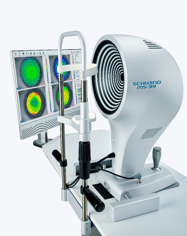
SCHWIND MS-39

We make the diagnosis with corneal tomography on the MS-39 AL. The device measures the front and back surfaces of the cornea, its thickness and the thickness of the outermost cell layer (epithelium). In early keratoconus in particular, the epithelium often shows typical changes.

Progression: when keratoconus is considered progressive

Whether keratoconus is progressing only becomes clear by comparing two measurements taken a few months apart. According to the global consensus, it is considered progressive if at least two values worsen: the front surface becomes steeper, the back surface becomes steeper, or the cornea becomes thinner (Gomes et al. 2015). The consensus does not specify a fixed number. This measurement determines whether crosslinking is performed and whether costs are covered.

Stages

Stages of keratoconus according to Amsler-Krumeich (simplified)
Stage Findings What usually helps
1 slight bulging, glasses or a soft contact lens are usually sufficient check-ups; crosslinking if it progresses
2 marked astigmatism, cornea still more than 400 µm thick, no scars crosslinking if it progresses, rigid contact lenses, topography-guided laser
3 pronounced bulging, cornea 300–400 µm CAIRS, crosslinking with an adapted protocol, special lenses
4 glasses prescription no longer measurable, central scars, very thin cornea special lenses, corneal transplant

The classification goes back to Krumeich, Daniel and Knülle 1998; the limits may differ in individual cases. What matters is not the stage alone, but whether the keratoconus is progressing.

Keratoconus in children, PMD and ectasia after LASIK

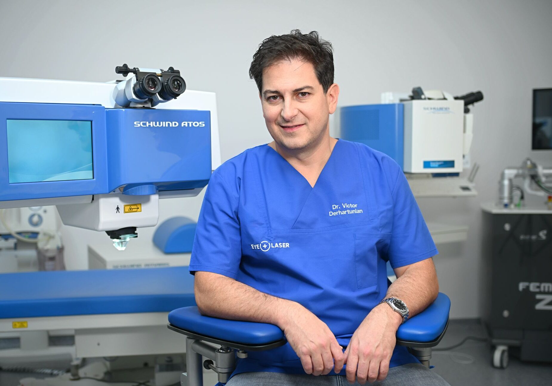
Dr. Victor Derhartunian

Keratoconus in children and adolescents

The younger the patient, the faster keratoconus often progresses. In a Zurich study with 59 eyes of 9- to 19-year-olds, it progressed in 88 %; the authors recommend not waiting for proof of progression before performing crosslinking in children (Chatzis and Hafezi 2012). We therefore check young patients at short intervals and discuss crosslinking with the parents early on. Important in everyday life: avoid rubbing the eyes and treat allergies.

Pellucid marginal degeneration (PMD)

PMD is a related condition: the cornea becomes thin not in the centre but in a narrow band at the lower edge. This causes pronounced astigmatism, often more than 5 dioptres. It usually appears later than keratoconus, at an average age of around 34, and more often in men (Sridhar et al. 2004). Treatment follows the same stages: crosslinking if it progresses, contact lenses, and ring segments in selected cases.

Ectasia after LASIK

Ectasia is a bulging of the cornea after a laser procedure, similar to keratoconus. It is rare: in an analysis of more than 30’000 LASIK eyes, it occurred in 0.033 % (Bohac et al. 2018). The most common risk factor is a cornea that was already irregular beforehand (Randleman et al. 2008); that is why we measure the cornea with tomography before every laser eye procedure. Ectasia is treated like progressive keratoconus, first with crosslinking. We will also examine you if you had your laser treatment elsewhere.

Who treats keratoconus at EyeLaser Zürich?

Dr. Victor Derhartunian

Crosslinking, CAIRS and all laser procedures are performed personally by Dr. med. univ. Victor Derhartunian, FEBO, FWCRS. He has been performing crosslinking since 2007 and was one of the first users in Europe after CE approval. He completed his specialist training in Erlangen and Frankfurt; he then worked at IROC in Zurich, where crosslinking was co-developed and clinically studied with Professor Theo Seiler.

«With crosslinking, we can halt progression in most cases. After that, the aim is to regain as much visual quality as possible.» — Dr. Victor Derhartunian

More about Dr. Victor Derhartunian

Our treatment approach: Stabilisation and rehabilitation

Our treatment plan for keratoconus is clearly structured and pursues two main goals. We explain every step to you transparently and in an easy-to-understand way.

Important note on objectivity: An excellent non-surgical option to improve vision is wearing rigid gas-permeable (hard) contact lenses. They compensate for the irregular corneal surface and often enable excellent vision. The following surgical procedures are primarily a solution for patients who cannot tolerate contact lenses or who want a permanent correction.

Step 1: Stabilising the tissue—the foundation

The most important goal of all is to stop the progression of keratoconus. For this, we use the gold standard:

  • Corneal crosslinking (CXL): This minimally invasive procedure strengthens the cornea through the combined use of vitamin B2 drops and UVA light. It makes the cornea stiffer and more resilient to prevent further bulging. In studies, crosslinking halted progression in around 92 to more than 98 out of 100 eyes: in a Zurich study with 117 eyes, keratoconus progressed further in 7.6 % after one year (Koller et al. 2009), and in a Dresden series with 241 eyes, two patients had to be treated again (Raiskup-Wolf et al. 2008).
Step 2: Visual rehabilitation—for the best possible vision

Once the disease has been stopped, we focus on improving your vision. It is important to understand that crosslinking alone rarely improves vision. True rehabilitation is achieved through modern, often combined procedures.

We offer the following treatment options at EyeLaser:

Phake Linsen (ICL) in Zürich
  • Combination of crosslinking and topography-guided Trans-PRK — more on this under Improving vision with keratoconus
    This modern method is one of our areas of focus. The goal is not only stabilisation but also the regularisation of the corneal surface. With topography-guided laser treatment, we smooth the irregular cornea in a targeted way. This reduces disruptive optical errors (higher-order aberrations) and can significantly improve visual quality with and without glasses.
Glaskörpertrübung im Auge
  • Modern biological ring segments: CAIRS (Corneal Allogenic Intrastromal Ring Segments)
    Instead of artificial plastic implants (PMMA), at EyeLaser we use natural donor tissue: CAIRS. These are precisely shaped segments made from human donor corneal tissue. The tissue contains no living donor cells; according to the studies to date, rejection and extrusion are therefore rare. Long-term data over many years is still lacking. The CAIRS integrate fully into your own cornea, strengthen it structurally and gently reshape it to reduce the bulging and improve visual quality.
  • Implantable contact lenses (ICL) for stable keratoconus
    After the cornea has been stabilised and regularised, residual refractive error may remain. If this is significant and the anatomy of the eye allows it, an implantable contact lens (ICL) can be inserted to achieve a high degree of independence from glasses.

Clinical example: Treating a complex case

Patient lying under the operating microscope while the surgeon in cap and mask leans over her

To illustrate the possibilities, here is an example of a multi-stage treatment plan for a very advanced keratoconus case:

  • Step 1: Implantation of CAIRS to mechanically reduce extreme corneal curvature.
  • Step 2 (after stabilisation): Performing a combined topo-guided Trans-PRK with crosslinking to further smooth the corneal surface and stop the disease.
  • Step 3 (after further healing): If a high residual refractive error remains, implantation of an ICL lens for final vision correction.

Overview

What treatments are there for keratoconus?

Keratoconus is treated in stages. The first goal is to stop it from progressing, and then to regain visual acuity. The overview shows what each procedure can and cannot do.

Keratoconus treatments compared — EyeLaser Zürich
Procedure Goal When it makes sense Stops progression Improves visual acuity Costs and insurance
Rigid contact lenses Visual acuity without a procedure as long as they are well tolerated no yes, while they are worn Fitting by a contact lens specialist
Crosslinking (CXL) Cross-link and stabilise the cornea when progression has been confirmed; in children often at diagnosis yes, in most cases usually only slightly, and that is not the goal Basic health insurance for progressive keratoconus; self-pay CHF 2’300 per eye
Topography-guided laser Smooth the corneal surface with a stable cornea that is not too thin, often together with crosslinking only together with crosslinking yes, especially visual quality CHF 2’500 per eye, self-pay; check cost approval
CAIRS Flatten the cornea with segments of donor tissue for more advanced deformation, when contact lenses are no longer sufficient no, usually combined with crosslinking yes CHF 4’500 per eye; cost approval in individual cases
ICL Correct remaining short-sightedness with stabilised, central keratoconus no yes, without glasses CHF 4’500 per eye, self-pay
Corneal transplant Replace diseased tissue when all gentler options have been exhausted yes yes, after a long healing period Basic health insurance; referral to a cornea centre

Two points that are often confused: Crosslinking stabilises the cornea; as a rule, it does not make your vision sharper; the improvement in vision comes from a second step. And crosslinking for progressive keratoconus is a mandatory benefit of basic health insurance (KLV Annex 1). We recommend obtaining a cost approval beforehand and will support you with this. All prices: Keratoconus costs.

The traffic-light system: Who is a suitable candidate for treatment?

Alterssichtigkeit behandeln in Zürich

🟢 Green light:

  • Patients with documented progression, typically under 40 years of age; children and adolescents often at diagnosis.
  • Sufficient corneal thickness for a safe CXL procedure.
  • No central corneal scarring.


🟡 Yellow light:

  • Patients with a thin cornea or for whom special protocols are required.
  • Patients over 40 with slow progression.
  • Pregnant or breastfeeding women (treatment is postponed).


🔴 Red light:

  • Patients with keratoconus that is too advanced, pronounced scarring or a cornea that is too thin.
  • Alternatives: Rigid contact lenses or, as a last resort, a corneal transplant (keratoplasty).

What happens next?

If keratoconus is suspected or for a follow-up check, make an appointment for an examination at Strehlgasse 33 in Zurich. Bring previous measurements and glasses prescriptions with you; the comparison shows whether the keratoconus is progressing. If there is a medical indication, we bill your health insurance under TARDOC.

Ask via WhatsApp Call 044 221 95 02

Online appointment

FAQ: Frequently asked questions

How can the progression of the disease be stopped?

The gold standard for treating progressive keratoconus is crosslinking (CXL), a procedure that strengthens the corneal structure and, in most cases, stops further progression of the disease.

How can vision be improved after stabilisation?

After crosslinking, modern procedures such as special laser eye treatments, biological ring segments or implantable lenses can regularise the cornea and significantly improve visual quality.

Who performs crosslinking in Zurich?

At EyeLaser Zürich, Dr. Victor Derhartunian performs crosslinking personally. He has been using the procedure since 2007 and was one of the first users in Europe after CE approval. He later worked at IROC in Zurich, where crosslinking was co-developed and studied with Professor Theo Seiler.

What non-surgical alternatives are available?

For many patients, rigid (hard) contact lenses are an excellent non-surgical option for achieving good vision. Surgical improvements are mainly an option when lenses are not tolerated.

What exactly is keratoconus?

Keratoconus is a condition in which the cornea becomes thinner and deforms into a cone shape, leading to irregular astigmatism and worsening vision.

What are the first symptoms?

Common early symptoms include blurred vision, rapid deterioration of eyesight, double vision in one eye and increased sensitivity to light.

Is keratoconus hereditary?

Yes, there is a genetic component. If someone in your family has keratoconus, your risk is increased.

Is eye rubbing really harmful?

Yes, definitely. Chronic eye rubbing is a major risk factor that can trigger or accelerate the disease.

Is the crosslinking treatment painful?

The procedure itself is painless thanks to anaesthetic drops. In the first 1–3 days afterwards, pain may occur, which is well controlled with medication.

Does health insurance in Switzerland cover the costs?

In Switzerland, crosslinking for progressive keratoconus is a mandatory benefit of basic health insurance (KLV Annex 1); the deductible and co-payment apply. EyeLaser Zürich recommends obtaining a cost approval beforehand and compiles the documents for you. As a self-payer, crosslinking costs CHF 2’300 per eye. For CAIRS, we clarify coverage on a case-by-case basis; you usually pay for ICL and laser treatment to improve vision yourself.

Will crosslinking alone improve my vision?

In most cases, the main goal of crosslinking is stabilisation, not improving vision. A significant improvement in visual quality is almost always achieved through additional procedures such as topography-guided laser treatment.

What are CAIRS and what is the advantage over other rings?

CAIRS are segments made from human donor corneal tissue, not plastic. Their excellent biocompatibility minimises the risk of complications such as rejection or inflammation compared with artificial implants.

Am I obliged to have surgery?

No, certainly not. We advise you objectively. If you cope well with rigid contact lenses and achieve excellent vision with them, there is often no reason for further surgical procedures to improve vision.

Can keratoconus be cured?

Keratoconus cannot be cured, but it can usually be halted. Crosslinking stiffens the cornea; in a Zurich study with 117 eyes, it progressed further afterwards in 7.6 % (Koller et al. 2009), and in a Dresden series the cornea remained stable over ten years (Raiskup et al. 2015). At EyeLaser Zürich, CAIRS, laser or ICL then improve vision.

Can you drive with keratoconus?

Yes, if the legally required visual acuity is achieved, with glasses or contact lenses. Keratoconus itself does not prohibit driving; what counts is your vision. However, glare at night can be a problem with keratoconus. At EyeLaser Zürich, we measure your visual acuity at every check-up and tell you whether you meet the requirements. The minimum requirements for vision in Annex 1 of the Swiss Traffic Licensing Ordinance (VZV) are decisive.

Can keratoconus cause blindness?

Blindness in the sense of complete loss of sight is not to be expected with keratoconus. Untreated, however, visual acuity can drop so much that glasses and contact lenses are no longer sufficient and a corneal transplant becomes necessary. Crosslinking has reduced this need in long-term studies (Raiskup et al. 2015). At EyeLaser Zürich, we therefore carry out check-ups early and regularly.

Can keratoconus be treated with a laser?

Normal laser eye surgery for freedom from glasses is not possible with keratoconus because it would weaken the cornea further. With stable keratoconus and a cornea that is not too thin, a topography-guided surface laser is possible, usually together with crosslinking: it smooths the cornea and improves visual quality. EyeLaser Zürich decides whether this is possible for you after the tomography.

What is ectasia after LASIK?

Ectasia after LASIK is a bulging of the cornea after the laser procedure, similar to keratoconus. It is rare: in more than 30’000 LASIK eyes, it occurred in 0.033 % (Bohac et al. 2018). It is treated like progressive keratoconus, first with crosslinking. EyeLaser Zürich also examines patients who had their laser treatment elsewhere.

From what age is crosslinking possible in children?

Crosslinking is also possible in children and adolescents; in a Zurich study, the patients treated were 9 to 19 years old (Chatzis and Hafezi 2012). Because keratoconus often progresses rapidly at this age, at EyeLaser Zürich we do not wait long and discuss crosslinking with the parents early on. Check-ups take place at short intervals.

Sources
  1. Gomes JA, Tan D, Rapuano CJ et al. Global consensus on keratoconus and ectatic diseases. Cornea 2015;34:359–369. PubMed 25738235
  2. Godefrooij DA, de Wit GA, Uiterwaal CS et al. Age-specific Incidence and Prevalence of Keratoconus: A Nationwide Registration Study. Am J Ophthalmol 2017;175:169–172. PubMed 28039037
  3. Hashemi H, Heydarian S, Hooshmand E et al. The Prevalence and Risk Factors for Keratoconus: A Systematic Review and Meta-Analysis. Cornea 2020;39:263–270. PubMed 31498247
  4. Koller T, Mrochen M, Seiler T. Complication and failure rates after corneal crosslinking. J Cataract Refract Surg 2009;35:1358–1362. PubMed 19631120
  5. Raiskup-Wolf F, Hoyer A, Spoerl E et al. Collagen crosslinking with riboflavin and ultraviolet-A light in keratoconus: long-term results. J Cataract Refract Surg 2008;34:796–801. PubMed 18471635
  6. Raiskup F, Theuring A, Pillunat LE, Spoerl E. Corneal collagen crosslinking with riboflavin and ultraviolet-A light in progressive keratoconus: ten-year results. J Cataract Refract Surg 2015;41:41–46. PubMed 25532633
  7. Chatzis N, Hafezi F. Progression of keratoconus and efficacy of pediatric corneal collagen cross-linking in children and adolescents. J Refract Surg 2012;28:753–758. PubMed 23347367
  8. Sridhar MS, Mahesh S, Bansal AK et al. Pellucid marginal corneal degeneration. Ophthalmology 2004;111:1102–1107. PubMed 15177959
  9. Bohac M, Koncarevic M, Pasalic A et al. Incidence and Clinical Characteristics of Post LASIK Ectasia: A Review of over 30,000 LASIK Cases. Semin Ophthalmol 2018;33:869–877. PubMed 30359155
  10. Randleman JB, Woodward M, Lynn MJ, Stulting RD. Risk assessment for ectasia after corneal refractive surgery. Ophthalmology 2008;115:37–50. PubMed 17624434
  11. Krumeich JH, Daniel J, Knülle A. Live-epikeratophakia for keratoconus. J Cataract Refract Surg 1998;24:456–463. PubMed 9584238
  12. Federal Office of Public Health (FOPH). Health Insurance Benefits Ordinance (KLV), Annex 1, edition of 1 July 2026, section 6 Ophthalmology.